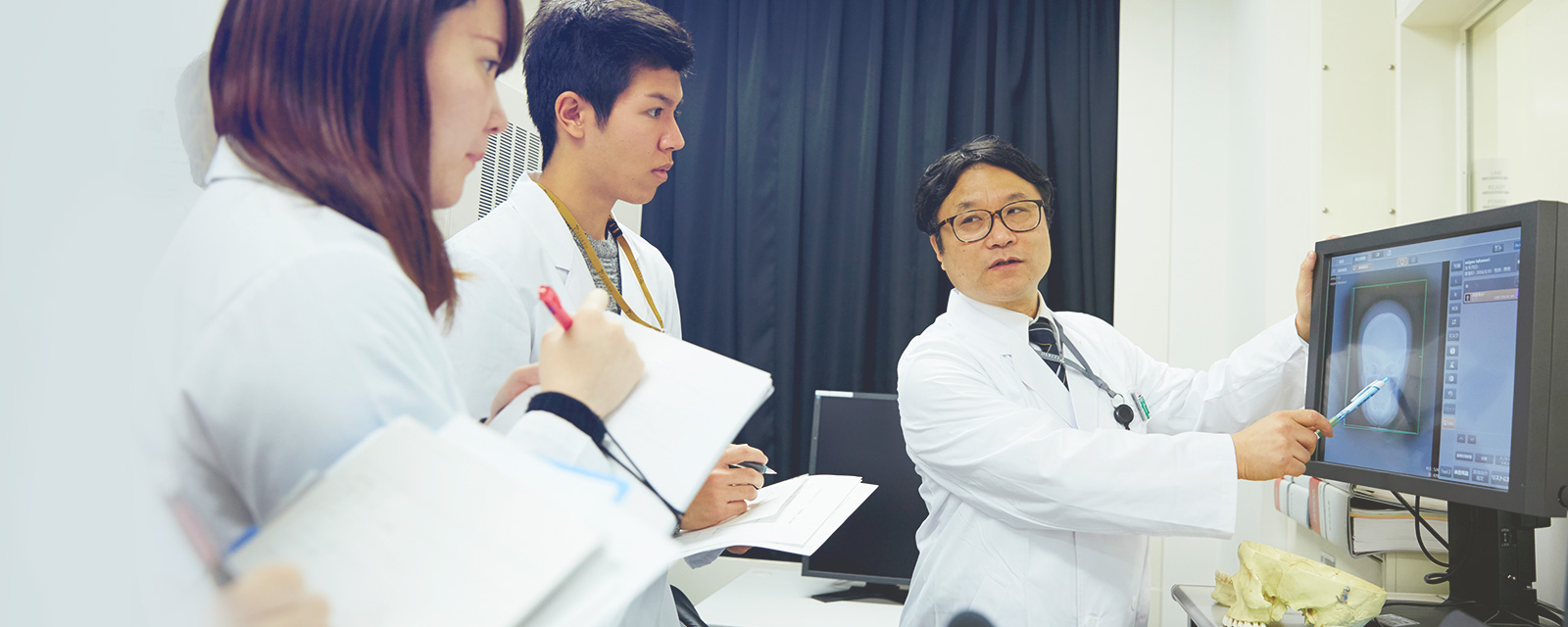
Educating specialists in radiology capable of responding to the advanced demands of clinical practice
?
?
Through in-depth guidance from faculty members in charge of each grade and a range of practical training programs, the Department of Radiological Technology provides students with the knowledge and skills they need to improve their practical skills. This is made possible by a well-developed learning environment; collaboration with the adjoining hospital of the School of Medicine, and advanced medical technology education provided by high-quality faculty members. In addition to the knowledge of radiation, the Department of Radiology trains radiological technologists who can play an active role in the rapidly changing medical field and contribute to society because they handle advanced and complex examination equipment and devices.
Department of Radiological Technology Close-Up
Grade level assignment system for in-depth instruction The Department has implemented a system of assigning teachers to each grade level that divides each grade level into small classes, with teachers assigned to each class in order to provide detailed instruction while maintaining communication. We aim to nurture a richer humanity with a high sense of ethics, knowledge, skills, and cultivation in our daily classes.
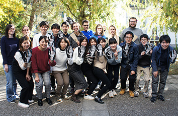
"Radiation Technology Training Overseas" The department offers an overseas training program in which students and faculty members can participate. At the Haute Ecole de Sante Vaud (HESAV), Canton Vaud, Switzerland, students receive practical training under the guidance of local doctors and radiological technologists, which is rarely experienced in Japan.
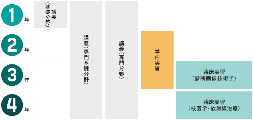
In the 1st year, students learn physics and chemistry experiments, which are the foundation of specialized subjects, and develop communication skills as medical professionals. In the 2nd year, students will acquire equipment operation and photography skills through lectures, experiments, and practical training, and in the 3rd year, clinical training at the adjoining hospital of the School of Medicine and other facilities will allow students to experience actual clinical settings and raise their awareness of the importance of being a medical professional. The final year of the program culminates in further clinical training, which consolidates the students' accumulated knowledge and experience. The students will increase their awareness as medical professionals and cultivate their abilities through comprehensive exercises and practical training.
e basis of specialized subjects, you will develop communication skills as a medical professional. After that, we will provide introductory education to specialized subjects and prepare for the 2nd year. In the 2nd year, you will acquire equipment operation and photography skills through lectures, experiments, and practical training. Through clinical training conducted at School of Medicine hospitals starting from the 3rd year, we will experience the actual field and raise awareness as a medical professional. After that, as a culmination of the final grade, we will work on further clinical training and consolidate the learning so far. Raise awareness as a medical professional and develop your skills through comprehensive exercises and practical training.
Radiation Physics I In this class, students first learn the basic structure of atoms, followed by the generation of X-rays and the interaction of X-rays with materials. Through the study of these fundamentals, students aim to acquire the basic skills needed to study specialized subjects such as "Radiation Physics II," "Medical Imaging Devices," and "Radiation Management."
Medical image informatics II (digital images) In this class, we will learn about the rapid shift to filmless technology in recent years and the "digital image processing" that is the mainstay of CAD (computer-aided diagnosis), which was approved by the U.S. FDA (Food Sanitation Administration) at the end of the 20th century. Digital image processing is part of the field of digital signal processing (DSP), so you should understand the basics of DSP and fast Fourier transform (FFT), learn sampling and quantization, mathematical models, image enhancement, etc. Learn the basics of clinical applications of digital image processing, and then learn about the basics of 3D medical imaging and CAD. By understanding this knowledge, we aim to improve our skills as specialists who will work in the medical imaging department in the future.
Radiation Therapy Technology I Although cancer is already a common problem in today's aging society, radiotherapy technology advancements are also impressive. In spite of the widely acknowledged risks associated with radiation exposure, radiotherapy technology is a study of the various justifications for using large doses of radiation on the human body. In this sense, it is fair to say that the most important thing about radiation therapy is to allow cancer cells to absorb an appropriate dose of radiation while protecting normal tissue as much as possible. The course aims to give students a thorough understanding of radiation therapy, including the interpretation of dose distribution, different types of radiation, the connection between the energy of radiation and the treatment site, knowledge of treatment equipment, and biology, leading to an understanding of the best radiation therapy for various types of cancers. We strive to develop staff members who, based on their understanding of radiation and cancer, can appreciate and manage the significance of the patient's emotional state and quality of life.
Experiment / Practice Field
Biomedical Engineering Experiments Most of the content of medical engineering is electrical and electronic engineering. We connect resistors, capacitors, coils, etc., pass signals through them, and observe what waveforms are produced. The purpose of this experiment is to find out whether or not the results match the theory we have learned so far, and if not, why not? 5 students work in pairs from preparation to report. In the past, we used a soldering iron to connect the components, but nowadays, thanks to the excellent breadboards, it is extremely easy to connect the components. In addition, there is a well-equipped facility with hundreds of components such as multimeters (which can measure voltage and capacitance) and other measuring instruments, function generators, and transistors. In addition to the lead professor, there are four or five teaching assistants (TAs) and auxiliary faculty members who follow up on experiments.
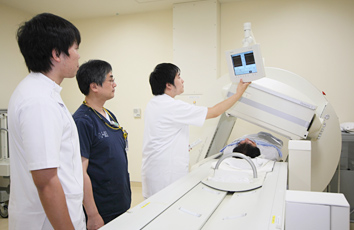
Basic Medical Imaging Technology Practice In the Basic Medical Imaging Technology Practice, students will learn basic experiments to study the characteristics and properties of X-rays, as well as how to use X-ray equipment, principles of operation, and various conditions necessary for X-ray imaging through hands-on experience. At the same time, students learn practical techniques and knowledge such as the fundamentals of radiographic reading that are linked to diagnostic imaging of various examinations; the mental attitude of a medical professional; methods of patient care; and countermeasures against infectious diseases.
Medical Imaging Technology Training I (Special Examination) In the Clinical Imaging Technology Training I (Special Examination), students will practice imaging techniques using special imaging devices. In gastrointestinal contrast examinations using FPDs, CR imaging, CT examinations, MRI examinations, and mammography examinations, students will take pictures of special phantoms (models) and various measuring tools, learning the principles and usage of the devices and how to evaluate their performance. In particular, in non-mydriatic fundus photography and ultrasound photography, which are not exposed to radiation, students will take turns as examiners and subjects and take pictures, aiming to acquire more practical skills and knowledge, including how to treat patients. In addition, an OSCE (Objective Structured Clinical Examination) will be held in the first semester, just before the hospital training.
Radiation management experiment In order to safely handle radiation and radioactive materials, it is natural that knowledge about radiation generating devices and radioactive isotopes is necessary, but from the perspective of "radiation management" it is important to know the dose limits for radiation exposure and avoid excessive exposure. Therefore, it is important to measure the concentration of radioactive substances in the environment to ensure safety. The purpose of this class is to learn how to measure the concentration of radioactive substances in various environments, and to experience the difficulty and importance of radiation measurement and management.
臨床実習
Training schedule
From the 3rd and 4th year years, clinical training will begin at medical institutions throughout the Kanto region, including the School of Medicine Hospital. By experientially learning the knowledge acquired in lectures and experiments in the actual field, we will surely acquire knowledge and skills. Through this highly specialized clinical training, you will develop your skills through comprehensive exercises and exams. You can also study abroad in Switzerland for a short period of time (conditions apply).
Clinical Training Subjects
Imaging diagnostic technology training (clinical training) photography edition This is my first practical training at a hospital. You will mainly learn about plain X-ray photography, X-ray fluoroscopic contrast examination (gastrointestinal tract, blood vessels), X-ray CT examination, MRI examination, mammography, bone density examination, and ultrasound examination. Hospital training is carried out at university hospitals, national and public hospitals, private general hospitals, etc., and teaches the role of radiological technologist in actual medical treatment, the necessity of Team Medical Care, patient treatment, X-ray photography technology, image examination technology, and more. will learn the manners of working adults. Before the hospital training, students receive detailed guidance from radiological technologist instructors, and after the hospital training, students present the results they have acquired during the clinical training at a clinical training report meeting. Department of Radiological Technology hopes that through hospital training, students will acquire Practical learning that cannot be learned through textbooks or on-campus lectures, and that they will increase their aspirations to radiological technologist.
Imaging diagnostic technology training (clinical training) OSCE edition Clinical training begins in the second half of your 3rd year, and is your first experience working with actual patients. Your activities, including observation, will be under the guidance and instructions of the person in charge of clinical training, but the on-campus X-ray photography training uses a phantom (model), so comprehensive skills including patient handling are required. Masu. The OSCE (Objective Structured Clinical Examination) is being implemented to fill this gap as much as possible. Specifically, the role of the patient is determined, and the trainee performs a series of practical tasks from start to finish, including taking pictures as instructed within a certain period of time, serving and guiding the patient (however, no X-rays are emitted), and then undergoes a test. It is evaluated by the instructor as follows. Not only will you get used to actually giving instructions to others, but by working seriously in the lead up to the exam, you will be better prepared, your knowledge will expand, and even better, as the instructors point out problems, you will be able to improve your own skills before clinical training. We aim to improve our shortcomings.
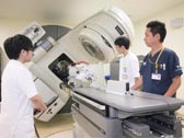
Nuclear medicine/radiation therapy training (clinical training) Nuclear medicine testing and radiation therapy fields both require large-scale equipment and equipment, so students will practice at clinical sites at external medical institutions based on the specialized knowledge they have learned from lectures within the university. Nuclear medicine testing involves injecting unsealed radioisotopes (RI) into the body as radiopharmaceuticals and detecting gamma rays emitted outside the body to diagnose disease states. You will also learn about radiation management. On the other hand, radiotherapy, which has recently attracted attention as a cancer treatment, uses high-energy radiation with a high voltage of over 1 million volts, unlike diagnostic X-rays. Therefore, it is of utmost importance to ensure accurate irradiation of the lesion, dose measurement, and quality assurance. It is also a rapidly evolving field, and provides a place to learn about new irradiation techniques. In both fields, patients who already have a disease are the main patients, so in addition to learning techniques and testing methods, you will also learn how to interact with patients, which can only be experienced in clinical settings.
List of main training destinations
Teikyo University Hospital, Teikyo University Hospital, Mizonokuchi, Tokyo Jiekai Medical University Hospital, Nippon Medical School Hospital, Showa University Hospital, Tokyo Women's Medical University Hospital, Keio University Hospital, Toranomon Hospital, JCHO Tokyo Shinjuku Medical Center, St. Luke's International Hospital, Nippon School of Medicine Itabashi Hospital, St. Marianna Medical University Hospital, Kanto Labor Disaster Hospital, National Cancer Research Center East Hospital, and others.
(As of April 2024)
成績評価と単位認定
Grading Criteria
Criteria For Advance to the next grade and Graduation certification, etc.
Annual promotion conditions and graduation / completion requirements are clearly stated in the course requirements, and are thoroughly known to students in the guidance at the beginning of the academic year. Advance to the next grade and graduation assessment meeting is held at the end of the year, and these are strictly operated based on the assessment materials. Failure to meet the requirements for advancement and graduation as specified in the course requirements results in retention in the original class.? The evaluation criteria for all subjects is specified in the course requirements distributed at the beginning of each semester. The evaluation scale varies depending on the subject, but in general, the grades of regular exams, grades of submissions such as reports, attendance status, and attitude of learning are apportioned and evaluated as a total.
Display of Grades and Assessment Criteria
Classification
Grading Criteria
GPA
Grading Criteria
Details of Assessment
Pass
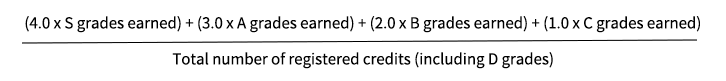
S.
4.0
90 percent or higher
Represents particularly excellent grades.
A
3.0
80 percent
Represents excellent grades.
B.
2.0
70 percent
Represents grades recognized as adequate.
C.
1.0
60 percent
Represents the minimum grade acceptable as a pass.
Fail
D.
0.0
Less than 60 percent
This means that the student has not reached the minimum grade acceptable as a pass. It also includes the lack of class attendance, the fact that the exams for the class have not been taken, and so on.
* GP: Points used to calculate GPA
About our GPA System
GPA (Grade Point Average) is a system that evaluates achievements of learning with objective numerical values. This system is generally based on the grade evaluation system based on universities in the United States and Europe.
GPA Calculation Method
Credit Recognition
Minimum number of courses or credits required for graduation
16 credits for compulsory subjects and 4 credits or more for elective subjects in basic field subjects, 20 credits or more in total, 34 credits for compulsory subjects in specialized basic field subjects, 70 credits for compulsory subjects in specialized field subjects, total 124 credits or more Must be.